Written by ashah-admin

Heart attack

A heart attack occurs when the flow of blood to the heart is blocked, most often by a build-up of fat, cholesterol and other substances, which form a plaque in the arteries that feed the heart (coronary arteries). The interrupted blood flow can damage or destroy part of the heart muscle.

A heart attack, also called a myocardial infarction, can be fatal, but treatment has improved dramatically over the years. It’s crucial to call 911 or emergency medical help if you think you might be having a heart attack.

Symptoms

Common heart attack signs and symptoms include:

  • Pressure, tightness, pain, or a squeezing or aching sensation in your chest or arms that may spread to your neck, jaw or back
  • Nausea, indigestion, heartburn or abdominal pain
  • Shortness of breath
  • Cold sweat
  • Fatigue
  • Lightheadedness or sudden dizziness

Heart attack symptoms vary

Not all people who have heart attacks have the same symptoms or have the same severity of symptoms. Some people have mild pain; others have more severe pain. Some people have no symptoms, while for others, the first sign may be sudden cardiac arrest. However, the more signs and symptoms you have, the greater the likelihood you’re having a heart attack.

Some heart attacks strike suddenly, but many people have warning signs and symptoms hours, days or weeks in advance. The earliest warning may be recurrent chest pain (angina) that’s triggered by exertion and relieved by rest. Angina is caused by a temporary decrease in blood flow to the heart.

A heart attack differs from a condition in which your heart suddenly stops (sudden cardiac arrest, which occurs when an electrical disturbance disrupts your heart’s pumping action and causes blood to stop flowing to the rest of your body). A heart attack can cause cardiac arrest, but it’s not the only cause.

Causes

A heart attack occurs when one or more of your coronary arteries become blocked. Over time, a coronary artery can narrow from the buildup of various substances, including cholesterol (atherosclerosis). This condition, known as coronary artery disease, causes most heart attacks.

During a heart attack, one of these plaques can rupture and spill cholesterol and other substances into the bloodstream. A blood clot forms at the site of the rupture. If large enough, the clot can completely block the flow of blood through the coronary artery.

Another cause of a heart attack is a spasm of a coronary artery that shuts down blood flow to part of the heart muscle. Use of tobacco and of illicit drugs, such as cocaine, can cause a life-threatening spasm. A heart attack can also occur due to a tear in the heart artery (spontaneous coronary artery dissection).

Treatments and drugs

Heart attack treatment at a hospital

With each passing minute after a heart attack, more heart tissue loses oxygen and deteriorates or dies. The main way to prevent heart damage is to restore blood flow quickly.

Medications

Medications given to treat a heart attack include:

  • Aspirin. The 911 operator may instruct you to take aspirin, or emergency medical personnel may give you aspirin immediately. Aspirin reduces blood clotting, thus helping maintain blood flow through a narrowed artery.
  • Thrombolytics. These drugs, also called clotbusters, help dissolve a blood clot that’s blocking blood flow to your heart. The earlier you receive a thrombolytic drug after a heart attack, the greater the chance you’ll survive and with less heart damage.
  • Antiplatelet agents. Emergency room doctors may give you other drugs to help prevent new clots and keep existing clots from getting larger. These include medications, such as clopidogrel (Plavix) and others, called platelet aggregation inhibitors.
  • Other blood-thinning medications. You’ll likely be given other medications, such as heparin, to make your blood less “sticky” and less likely to form clots. Heparin is given intravenously or by an injection under your skin.
  • Pain relievers. You may receive a pain reliever, such as morphine, to ease your discomfort.
  • Nitroglycerin. This medication, used to treat chest pain (angina), can help improve blood flow to the heart by widening (dilating) the blood vessels.
  • Beta blockers. These medications help relax your heart muscle, slow your heartbeat and decrease blood pressure, making your heart’s job easier. Beta blockers can limit the amount of heart muscle damage and prevent future heart attacks.
  • ACE inhibitors. These drugs lower blood pressure and reduce stress on the heart.
Surgical and other procedures

In addition to medications, you may undergo one of the following procedures to treat your heart attack:

  • Coronary angioplasty and stenting. Doctors insert a long, thin tube (catheter) that’s passed through an artery, usually in your leg or groin, to a blocked artery in your heart. If you’ve had a heart attack, this procedure is often done immediately after a cardiac catheterization, a procedure used to locate blockages. This catheter is equipped with a special balloon that, once in position, is briefly inflated to open a blocked coronary artery. A metal mesh stent may be inserted into the artery to keep it open long term, restoring blood flow to the heart. Depending on your condition, your doctor may opt to place a stent coated with a slow-releasing medication to help keep your artery open.
  • Coronary artery bypass surgery. In some cases, doctors may perform emergency bypass surgery at the time of a heart attack. If possible, your doctor may suggest that you have bypass surgery after your heart has had time — about three to seven days — to recover from your heart attack.
    Bypass surgery involves sewing veins or arteries in place beyond a blocked or narrowed coronary artery, allowing blood flow to the heart to bypass the narrowed section.

Once blood flow to your heart is restored and your condition is stable, you’re likely to remain in the hospital for several days.

Prevention

It’s never too late to take steps to prevent a heart attack — even if you’ve already had one. Here are ways to prevent a heart attack.

  • Medications. Taking medications can reduce your risk of a subsequent heart attack
    and help your damaged heart function better. Continue to take what your doctor
    prescribes, and ask your doctor how often you need to be monitored.
  • Lifestyle factors. You know the drill: Maintain a healthy weight with a heart-healthy
    diet, don’t smoke, exercise regularly, manage stress and control conditions that can
    lead to heart attack, such as high blood pressure, high cholesterol and diabetes.
Written by ashah-admin

Gangrene

Gangrene refers to the death of body tissue due to a lack of blood flow or a bacterial infection. Gangrene most commonly affects the extremities, including your toes, fingers and limbs, but it can also occur in your muscles and internal organs.

Your chances of developing gangrene are higher if you have an underlying condition that can damage your blood vessels and affect blood flow, such as diabetes or atherosclerosis.

Treatments for gangrene include surgery to remove dead tissue, antibiotics and other approaches. The prognosis for recovery is better if gangrene is identified early and treated quickly.

Symptoms

When gangrene affects your skin, signs and symptoms may include:

  • Skin discoloration — ranging from pale to blue, purple, black, bronze or red, depending on the type of gangrene you have
  • A clear line between healthy and damaged skin
  • Severe pain followed by a feeling of numbness
  • A foul-smelling discharge leaking from a sore

If you have a type of gangrene that affects tissues beneath the surface of your skin, such as gas gangrene or internal gangrene, you may notice that:

  • The affected tissue is swollen and very painful
  • You’re running a fever and feel unwell

A condition called septic shock can occur if a bacterial infection that originated in the gangrenous tissue spreads throughout your body. Signs and symptoms of septic shock include:

  • Low blood pressure
  • Fever, possibly, though temperature may also run lower than the normal 96.8 F (36 C)
  • Rapid heart rate
  • Lightheadedness
  • Shortness of breath
  • Confusion

Causes

Gangrene may occur due to one or both of the following:

  • Lack of blood supply. Your blood provides oxygen, nutrients to feed your cells, and immune system components, such as antibodies, to ward off infections. Without a proper blood supply, cells can’t survive, and your tissue decays.
  • Infection. If bacteria thrive unchecked for long, infection can take over and cause your tissue to die, causing gangrene.

Types of gangrene

    • Dry gangrene. Dry gangrene is characterized by dry and shriveled skin ranging in color from brown to purplish-blue to black. Dry gangrene may develop slowly. It occurs most commonly in people who have a blood vessel disease, such as atherosclerosis.
    • Wet gangrene. Gangrene is referred to as “wet” if there’s a bacterial infection in the affected tissue. Swelling, blistering and a wet appearance are common features of wet gangrene.It may develop after a severe burn, frostbite or injury. It often occurs in people with diabetes who unknowingly injure a toe or foot. Wet gangrene needs to be treated immediately because it spreads quickly and can be fatal.
    • Gas gangrene. Gas gangrene typically affects deep muscle tissue. If you have gas gangrene, the surface of your skin may initially appear normal.As the condition progresses, your skin may become pale and then evolve to a gray or purplish-red color. A bubbly appearance to your skin may become apparent, and the affected skin may make a crackling sound when you press on it because of the gas within the tissue.Gas gangrene is commonly caused by infection with the bacterium Clostridium perfringens, which develops in an injury or surgical wound that’s depleted of blood supply. The bacterial infection produces toxins that release gas — hence the name “gas” gangrene — and cause tissue death. Like wet gangrene, gas gangrene can be life-threatening.

<li>Internal gangrene. Gangrene affecting one or more of your organs, such as your intestines, gallbladder or appendix, is called internal gangrene. This type of gangrene occurs when blood flow to an internal organ is blocked — for example, when your intestines bulge through a weakened area of muscle in your abdomen (hernia) and become twisted.

Internal gangrene may cause fever and severe pain. Left untreated, internal gangrene can be fatal.

  • Fournier’s gangrene. Fournier’s gangrene involves the genital organs. Men are more often affected, but women can develop this type of gangrene as well. Fournier’s gangrene usually arises due to an infection in the genital area or urinary tract and causes genital pain, tenderness, redness and swelling.
  • Progressive bacterial synergistic gangrene (Meleney’s gangrene). This rare type of gangrene typically occurs after an operation, with painful skin lesions developing one to two weeks after surgery.


Treatments and drugs

Tissue that has been damaged by gangrene can’t be saved, but steps can be taken to prevent gangrene from progressing. These treatments include:

  • Surgery. Your doctor removes the dead tissue, which helps stop gangrene from spreading and allows healthy tissue to heal. If possible, your doctor may repair damaged or diseased blood vessels in order to increase blood flow to the affected area.A skin graft is a type of reconstructive surgery that may be used to repair damage to your skin caused by gangrene. During a skin graft, your doctor removes healthy skin from another part of your body — usually a place hidden by clothing — and carefully spreads it over an affected area.The healthy skin may be held in place by a dressing or by a couple of small stitches. A skin graft can be done only if an adequate blood supply has been restored to the damaged skin.In severe cases of gangrene, an affected body part, such as a toe, finger or limb, may need to be surgically removed (amputated). In some cases, you may later be fitted with an artificial limb (prosthesis).
  • Antibiotics. Antibiotics given through a vein (intravenous) may be used to treat gangrene that’s become infected.
  • Hyperbaric oxygen therapy. In addition to antibiotics and surgery, hyperbaric oxygen therapy also may be used to treat gangrene. Under increased pressure and increased oxygen content, your blood is able to carry greater amounts of oxygen. Blood rich in oxygen slows the growth of bacteria that thrive in the absence of oxygen and helps infected wounds heal more easily.In this type of therapy, you’ll be situated in a special chamber, which usually consists of a padded table that slides into a clear plastic tube. The chamber is pressurized with pure oxygen, and the pressure inside the chamber will slowly rise to about two and a half times normal atmospheric pressure.Hyperbaric oxygen therapy for gas gangrene generally lasts about 90 minutes. You may need two to three treatments daily.

Other treatments for gangrene may include supportive care, including fluids, nutrients and pain medication to relieve your discomfort.

Outlook

Generally, people who have dry gangrene have the best chance of a full recovery because dry gangrene doesn’t involve a bacterial infection and spreads more slowly than do the other types of gangrene. However, when gangrene caused by an infection is recognized and treated quickly, the odds of recovery are good.

Prevention

Here are a few suggestions to help you reduce your risk of developing gangrene:

  • Care for your diabetes. If you have diabetes, make sure you examine your hands and feet daily for cuts, sores and signs of infection, such as redness, swelling or drainage. Ask your doctor to examine your hands and feet at least once a year.
  • Lose weight. Excess pounds not only put you at risk of diabetes but also place pressure on your arteries, constricting blood flow and putting you at risk of infection and slow wound healing.
  • Don’t use tobacco. The chronic use of tobacco products can damage your blood vessels.
  • Help prevent infections. Wash any open wounds with a mild soap and water and try to keep them clean and dry until they heal.
  • Watch out when the temperature drops. Frostbitten skin can lead to gangrene because frostbite reduces blood circulation in an affected area. If you notice that any area of your skin has become pale, hard, cold and numb after prolonged exposure to cold temperatures, call your doctor.
Written by ashah-admin

Frozen shoulder

Frozen shoulder, also known as adhesive capsulitis, is a condition characterized by stiffness and pain in your shoulder joint. Signs and symptoms typically begin gradually, worsen over time and then resolve, usually within one to three years.

Your risk of developing frozen shoulder increases if you’re recovering from a medical condition or procedure that prevents you from moving your arm — such as a stroke or a mastectomy.

Treatment for frozen shoulder involves range-of-motion exercises and, sometimes, corticosteroids and numbing medications injected into the joint capsule. In a small percentage of cases, arthroscopic surgery may be indicated to loosen the joint capsule so that it can move more freely. It’s unusual for frozen shoulder to recur in the same shoulder, but some people can develop it in the opposite shoulder.

Symptoms

Frozen shoulder typically develops slowly, and in three stages. Each stage can last a number of months.

  • Freezing stage. Any movement of your shoulder causes pain, and your shoulder’s range of motion starts to become limited.
  • Frozen stage. Pain may begin to diminish during this stage. However, your shoulder becomes stiffer, and using it becomes more difficult.
  • Thawing stage. The range of motion in your shoulder begins to improve.

For some people, the pain worsens at night, sometimes disrupting sleep.

Causes

The bones, ligaments and tendons that make up your shoulder joint are encased in a capsule of connective tissue. Frozen shoulder occurs when this capsule thickens and tightens around the shoulder joint, restricting its movement.

Doctors aren’t sure why this happens to some people, although it’s more likely to occur in people who have diabetes or those who recently had to immobilize their shoulder for a long period, such as after surgery or an arm fracture.

Treatments and drugs

Most frozen shoulder treatment involves controlling shoulder pain and preserving as much range of motion in the shoulder as possible.

Medications

Over-the-counter pain relievers, such as aspirin and ibuprofen (Advil, Motrin IB, others), can help reduce pain and inflammation associated with frozen shoulder. In some cases, your doctor may prescribe stronger pain-relieving and anti-inflammatory drugs.

Therapy

A physical therapist can teach you range-of-motion exercises to help recover as much mobility in your shoulder as possible. Your commitment to doing these exercises is important to optimize recovery of your mobility.

Surgical and other procedures

Most frozen shoulders get better on their own within 12 to 18 months. For persistent symptoms, your doctor may suggest:

  • Steroid injections. Injecting corticosteroids into your shoulder joint may help decrease pain and improve shoulder mobility, especially in the early stages of the process.
  • Joint distension. Injecting sterile water into the joint capsule can help stretch the tissue and make it easier to move the joint.
  • Shoulder manipulation. In this procedure, you receive a general anesthetic, so you’ll be unconscious and feel no pain. Then the doctor moves your shoulder joint in different directions, to help loosen the tightened tissue.
  • Surgery. Surgery for frozen shoulder is rare, but if nothing else has helped, your doctor may recommend surgery to remove scar tissue and adhesions from inside your shoulder joint. Doctors usually perform this surgery with lighted, tubular instruments inserted through small incisions around your joint (arthroscopically).

Prevention

  • One of the most common causes of frozen shoulder is the immobility that may result during recovery from a shoulder injury, broken arm or a stroke. If you’ve had an injury that makes it difficult to move your shoulder, talk to your doctor about exercises you can do to maintain the range of motion in your shoulder joint.
Written by ashah-admin

Diabetes

Diabetes mellitus refers to a group of diseases that affect how your body uses blood sugar (glucose). Glucose is vital to your health because it’s an important source of energy for the cells that make up your muscles and tissues. It’s also your brain’s main source of fuel.

If you have diabetes, no matter what type, it means you have too much glucose in your blood, although the causes may differ. Too much glucose can lead to serious health problems.

Chronic diabetes conditions include type 1 diabetes and type 2 diabetes. Potentially reversible diabetes conditions include prediabetes — when your blood sugar levels are higher than normal, but not high enough to be classified as diabetes — and gestational diabetes, which occurs during pregnancy but may resolve after the baby is delivered.

Symptoms

Diabetes symptoms vary depending on how much your blood sugar is elevated. Some people, especially those with prediabetes or type 2 diabetes, may not experience symptoms initially. In type 1 diabetes, symptoms tend to come on quickly and be more severe.

Some of the signs and symptoms of type 1 and type 2 diabetes are:

  • Increased thirst
  • Frequent urination
  • Extreme hunger
  • Unexplained weight loss
  • Presence of ketones in the urine (ketones are a byproduct of the breakdown of muscle and fat that happens when there’s not enough available insulin)
  • Fatigue
  • Irritability
  • Blurred vision
  • Slow-healing sores
  • Frequent infections, such as gums or skin infections and vaginal infections

Although type 1 diabetes can develop at any age, it typically appears during childhood or adolescence. Type 2 diabetes, the more common type, can develop at any age, though it’s more common in people older than 40.

Causes

To understand diabetes, first you must understand how glucose is normally processed in the body.

How insulin works

Insulin is a hormone that comes from a gland situated behind and below the stomach (pancreas).

  • The pancreas secretes insulin into the bloodstream.
  • The insulin circulates, enabling sugar to enter your cells.
  • Insulin lowers the amount of sugar in your bloodstream.
  • As your blood sugar level drops, so does the secretion of insulin from your pancreas

The role of glucose

Glucose — a sugar — is a source of energy for the cells that make up muscles and other tissues.

  • Glucose comes from two major sources: food and your liver.
  • Sugar is absorbed into the bloodstream, where it enters cells with the help of insulin.
  • Your liver stores and makes glucose.
  • When your glucose levels are low, such as when you haven’t eaten in a while, the liver breaks down stored glycogen into glucose to keep your glucose level within a normal range.

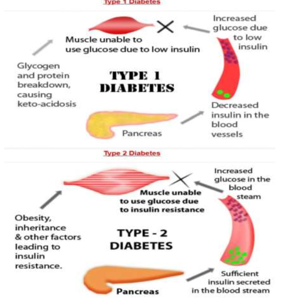
Causes of type 1 diabetes

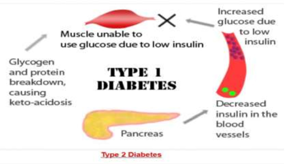
The exact cause of type 1 diabetes is unknown. What is known is that your immune system — which normally fights harmful bacteria or viruses — attacks and destroys your insulin-producing cells in the pancreas. This leaves you with little or no insulin. Instead of being transported into your cells, sugar builds up in your bloodstream.

Type 1 is thought to be caused by a combination of genetic susceptibility and environmental factors, though exactly what many of those factors are is still unclear.

Causes of prediabetes and type 2 diabetes

In prediabetes — which can lead to type 2 diabetes — and in type 2 diabetes, your cells become resistant to the action of insulin, and your pancreas is unable to make enough insulin to overcome this resistance. Instead of moving into your cells where it’s needed for energy, sugar builds up in your bloodstream.

Exactly why this happens is uncertain, although it’s believed that genetic and environmental factors play a role in the development of type 2 diabetes. Being overweight is strongly linked to the development of type 2 diabetes, but not everyone with type 2 is overweight.

Causes of gestational diabetes

During pregnancy, the placenta produces hormones to sustain your pregnancy. These hormones make your cells more resistant to insulin.

Normally, your pancreas responds by producing enough extra insulin to overcome this resistance. But sometimes your pancreas can’t keep up. When this happens, too little glucose gets into your cells and too much stays in your blood, resulting in gestational diabetes.

Treatments and drugs

Depending on what type of diabetes you have, blood sugar monitoring, insulin and oral medications may play a role in your treatment. Eating a healthy diet, maintaining a healthy weight and participating in regular activity also are important factors in managing diabetes.

Treatments for all types of diabetes

An important part of managing diabetes — as well as your overall health — is maintaining a healthy weight through a healthy diet and exercise plan:

  • Healthy eating. Contrary to popular perception, there’s no specific diabetes diet. You’ll need to center your diet on more fruits, vegetables and whole grains — foods that are high in nutrition and fiber and low in fat and calories — and cut down on animal products, refined carbohydrates and sweets. In fact, it’s the best eating plan for the entire family. Sugary foods are OK once in a while, as long as they’re counted as part of your meal plan.Yet understanding what and how much to eat can be a challenge. A registered dietitian can help you create a meal plan that fits your health goals, food preferences and lifestyle. This will likely include carbohydrate counting, especially if you have type 1 diabetes.
  • Physical activity. Everyone needs regular aerobic exercise, and people who have diabetes are no exception. Exercise lowers your blood sugar level by moving sugar into your cells, where it’s used for energy. Exercise also increases your sensitivity to insulin, which means your body needs less insulin to transport sugar to your cells. Get your doctor’s OK to exercise. Then choose activities you enjoy, such as walking, swimming or biking. What’s most important is making physical activity part of your daily routine. Aim for at least 30 minutes or more of aerobic exercise most days of the week. If you haven’t been active for a while, start slowly and build up gradually.

Treatments for type 1 and type 2 diabetes

Treatment for type 1 diabetes involves insulin injections or the use of an insulin pump, frequent blood sugar checks, and carbohydrate counting. Treatment of type 2 diabetes primarily involves monitoring of your blood sugar, along with diabetes medications, insulin or both.

  • Monitoring your blood sugar. Depending on your treatment plan, you may check and record your blood sugar as often as several times a week to as many as four to eight times a day. Careful monitoring is the only way to make sure that your blood sugar level remains within your target range. People who receive insulin therapy also may choose to monitor their blood sugar levels with a continuous glucose monitor. Although this technology doesn’t yet replace the glucose meter, it can provide important information about trends in blood sugar levels.Even with careful management, blood sugar levels can sometimes change unpredictably. With help from your diabetes treatment team, you’ll learn how your blood sugar level changes in response to food, physical activity, medications, illness, alcohol, stress — for women, fluctuations in hormone levels.In addition to daily blood sugar monitoring, your doctor will likely recommend regular A1C testing to measure your average blood sugar level for the past two to three months. Compared with repeated daily blood sugar tests, A1C testing better indicates how well your diabetes treatment plan is working overall. An elevated A1C level may signal the need for a change in your insulin regimen or meal plan. Your target A1C goal may vary depending on your age and various other factors. However, for most people with diabetes, the American Diabetes Association recommends an A1C of below 7 percent. Ask your doctor what your A1C target is.
  • Insulin. People with type 1 diabetes need insulin therapy to survive. Many people with type 2 diabetes or gestational diabetes also need insulin therapy.Many types of insulin are available, including rapid-acting insulin, long-acting insulin and intermediate options. Depending on your needs, your doctor may prescribe a mixture of insulin types to use throughout the day and night.Insulin can’t be taken orally to lower blood sugar because stomach enzymes interfere with insulin’s action. Often insulin is injected using a fine needle and syringe or an insulin pen — a device that looks like a large ink pen.An insulin pump may also be an option. The pump is a device about the size of a cellphone worn on the outside of your body. A tube connects the reservoir of insulin to a catheter that’s inserted under the skin of your abdomen. A tubeless pump that works wirelessly is also now available. You program an insulin pump to dispense specific amounts of insulin. It can be adjusted to deliver more or less insulin depending on meals, activity level and blood sugar level.

    An emerging treatment approach, not yet available, is closed loop insulin delivery, also known as the artificial pancreas. It links a continuous glucose monitor to an insulin pump. The device automatically delivers the correct amount of insulin when the monitor indicates the need for it. There are a number of different versions of the artificial pancreas, and clinical trials have had encouraging results. More research needs to be done before a fully functional artificial pancreas can receive regulatory approval.

    However, the first step toward an artificial pancreas was approved in 2013. Combining a continuous glucose monitor with an insulin pump, this system stops insulin delivery when blood sugar levels drop too low. Studies on the device found that it could prevent low blood sugar levels overnight without significantly increasing morning blood sugar levels.

  • Oral or other medications. Sometimes other oral or injected medications are prescribed as well. Some diabetes medications stimulate your pancreas to produce and release more insulin. Others inhibit the production and release of glucose from your liver, which means you need less insulin to transport sugar into your cells. Still others block the action of stomach or intestinal enzymes that break down carbohydrates or make your tissues more sensitive to insulin. Metformin (Glucophage, Glumetza, others) is generally the first medication prescribed for type 2 diabetes.
  • Transplantation. In some people who have type 1 diabetes, a pancreas transplant may be an option. Islet transplants are being studied as well. With a successful pancreas transplant, you would no longer need insulin therapy. But transplants aren’t always successful — and these procedures pose serious risks. You need a lifetime of immune-suppressing drugs to prevent organ rejection. These drugs can have serious side effects, including a high risk of infection, organ injury and cancer. Because the side effects can be more dangerous than the diabetes, transplants are usually reserved for people whose diabetes can’t be controlled or those who also need a kidney transplant.
  • Bariatric surgery. Although it is not specifically considered a treatment for type 2 diabetes, people with type 2 diabetes who also have a body mass index higher than 35 may benefit from this type of surgery. People who’ve undergone gastric bypass have seen significant improvements in their blood sugar levels. However, this procedure’s long-term risks and benefits for type 2 diabetes aren’t yet known.

Treatment for gestational diabetes

Controlling your blood sugar level is essential to keeping your baby healthy and avoiding complications during delivery. In addition to maintaining a healthy diet and exercising, your treatment plan may include monitoring your blood sugar and, in some cases, using insulin or oral medications.

Your health care provider will also monitor your blood sugar level during labor. If your blood sugar rises, your baby may release high levels of insulin — which can lead to low blood sugar right after birth.

Treatment for prediabetes

If you have prediabetes, healthy lifestyle choices can help you bring your blood sugar level back to normal or at least keep it from rising toward the levels seen in type 2 diabetes. Maintaining a healthy weight through exercise and healthy eating can help. Exercising at least 150 minutes a week and losing 5 to 10 percent of your body weight may prevent or delay type 2 diabetes.

Sometimes medications — such as metformin (Glucophage, Glumetza, others) — also are an option if you’re at high risk of diabetes, including when your prediabetes is worsening or if you have cardiovascular disease, fatty liver disease or polycystic ovary syndrome.

In other cases, medications to control cholesterol — statins, in particular — and high blood pressure medications are needed. Your doctor might prescribe low-dose aspirin therapy to help prevent cardiovascular disease if you’re at high risk. Healthy lifestyle choices remain key, however.

Signs of trouble in any type of diabetes

Because so many factors can affect your blood sugar, problems may sometimes arise that require immediate care, such as:

  • High blood sugar (hyperglycemia). Your blood sugar level can rise for many reasons, including eating too much, being sick or not taking enough glucoselowering medication. Check your blood sugar level as directed by your doctor, and watch for signs and symptoms of high blood sugar — frequent urination, increased thirst, dry mouth, blurred vision, fatigue and nausea. If you have hyperglycemia, you’ll need to adjust your meal plan, medications or both.
  • Increased ketones in your urine (diabetic ketoacidosis). If your cells are starved for energy, your body may begin to break down fat. This produces toxic acids known as ketones. Watch for loss of appetite, weakness, vomiting, fever, stomach pain and a sweet, fruity breath. You can check your urine for excess ketones with an over-thecounter ketones test kit. If you have excess ketones in your urine, consult your doctor right away or seek emergency care. This condition is more common in people with type 1 diabetes.
  • Hyperglycemic hyperosmolar nonketotic syndrome. Signs and symptoms of this life-threatening condition include a blood sugar reading over 600 mg/dL (33.3 mmol/L), dry mouth, extreme thirst, fever, drowsiness, confusion, vision loss and hallucinations. Hyperosmolar syndrome is caused by sky-high blood sugar that turns blood thick and syrupy. It tends to be more common in people with type 2 diabetes, and it’s often preceded by an illness. Call your doctor or seek immediate medical care if you have signs or symptoms of this condition.
  • Low blood sugar (hypoglycemia). If your blood sugar level drops below your target range, it’s known as low blood sugar (hypoglycemia). Your blood sugar level can drop for many reasons, including skipping a meal and getting more physical activity than normal. However, low blood sugar is most likely if you take glucoselowering medications that promote the secretion of insulin by your pancreas or if you’re receiving insulin therapy. Check your blood sugar level regularly, and watch for signs and symptoms of low blood sugar — sweating, shakiness, weakness, hunger, dizziness, headache, blurred vision, heart palpitations, irritability, slurred speech, drowsiness, confusion, fainting and seizures. Low blood sugar is treated with quickly absorbed carbohydrates, such as fruit juice or glucose tablets.

Prevention

Type 1 diabetes can’t be prevented. However, the same healthy lifestyle choices that help treat prediabetes, type 2 diabetes and gestational diabetes can also help prevent them:

  • Eat healthy foods. Choose foods lower in fat and calories and higher in fiber. Focus on fruits, vegetables and whole grains. Strive for variety to prevent boredom.
  • Get more physical activity. Aim for 30 minutes of moderate physical activity a day. Take a brisk daily walk. Ride your bike. Swim laps. If you can’t fit in a long workout, break it up into smaller sessions spread throughout the day.
  • Lose excess pounds. If you’re overweight, losing even 7 percent of your body weight — for example, 14 pounds (6.4 kilograms) if you weigh 200 pounds (90.9 kilograms) — can reduce the risk of diabetes. To keep your weight in a healthy range, focus on permanent changes to your eating and exercise habits. Motivate yourself by remembering the benefits of losing weight, such as a healthier heart, more energy and improved self-esteem.
  • Sometimes medication is an option as well. Oral diabetes drugs such as metformin (Glucophage, Glumetza, others) may reduce the risk of type 2 diabetes — but healthy lifestyle choices remain essential.
  • Have your blood sugar checked at least once a year to check that you haven’t developed type 2 diabetes.
Written by ashah-admin

Dandruff

Definition

Dandruff is a common chronic scalp condition marked by flaking of the skin on your scalp. Although dandruff isn’t contagious and is rarely serious, it can be embarrassing and sometimes difficult to treat.

The good news is that dandruff usually can be controlled. Mild cases of dandruff may need nothing more than daily shampooing with a gentle cleanser. More stubborn cases of dandruff often respond to medicated shampoos.

Symptoms

For most teens and adults, dandruff symptoms are easy to spot: white, oily looking flakes of dead skin that dot your hair and shoulders, and a possibly itchy, scaly scalp. The condition may worsen during the fall and winter, when indoor heating can contribute to dry skin, and improve during the summer.

A type of dandruff called cradle cap can affect babies. This disorder, which causes a scaly, crusty scalp, is most common in newborns, but it can occur anytime during infancy. Although it can be alarming for parents, cradle cap isn’t dangerous and usually clears up on its own by the time a baby is 3 years old.

Causes

Dandruff can have several causes, including:

  • Dry skin. Simple dry skin is the most common cause of dandruff. Flakes from dry skin are generally smaller and less oily than those from other causes of dandruff, and you’ll likely have symptoms and signs of dry skin on other parts of the body, such as your legs and arms.
  • Irritated, oily skin (seborrheic dermatitis). This condition, one of the most frequent causes of dandruff, is marked by red, greasy skin covered with flaky white or yellow scales. Seborrheic dermatitis may affect your scalp and other areas rich in oil glands, such as your eyebrows, the sides of your nose and the backs of your ears, your breastbone, your groin area, and sometimes your armpits.
  • Not shampooing often enough. If you don’t regularly wash your hair, oils and skin cells from your scalp can build up, causing dandruff.
  • Other skin conditions. People with skin conditions such as eczema — a chronic, inflammatory skin condition — or psoriasis — a skin condition marked by a rapid buildup of rough, dry, dead skin cells that form thick scales — may appear to have dandruff.
  • A yeast-like fungus (malassezia). Malassezia lives on the scalps of most adults, but for some, it irritates the scalp. This can irritate your scalp and cause more skin cells to grow. The extra skin cells die and fall off, making them appear white and flaky in your hair or on your clothes. Why malassezia irritates some scalps isn’t known.
  • Sensitivity to hair care products (contact dermatitis). Sometimes sensitivities to certain ingredients in hair care products or hair dyes, especially paraphenylenediamine, can cause a red, itchy, scaly scalp. Shampooing too often or using too many styling products also may irritate your scalp, causing dandruff.

Treatments and drugs

Dandruff can almost always be controlled, but dandruff treatment may take patience and persistence. In general, daily cleansing with a gentle shampoo to reduce oiliness and skin cell buildup can often help mild dandruff.

When regular shampoos fail, dandruff shampoos you can buy at a drugstore may succeed. But dandruff shampoos aren’t all alike, and you may need to experiment until you find one that works for you. If you develop itching, stinging, redness or burning from any product, stop using it. If you develop an allergic reaction, such as a rash, hives or difficulty breathing, seek immediate medical attention.

Dandruff shampoos are classified according to the medication they contain:

  • Zinc pyrithione shampoos (such as Head & Shoulders, Jason Dandruff Relief 2 in 1, others). These contain the antibacterial and antifungal agent zinc pyrithione, which can reduce the fungus on your scalp that can cause dandruff and seborrheic dermatitis.
  • Tar-based shampoos (such as Neutrogena T/Gel). Coal tar, a byproduct of the coal manufacturing process, helps conditions such as dandruff, seborrheic dermatitis and psoriasis by slowing how quickly skin cells on your scalp die and flake off.  Shampoos containing salicylic acid (such as Neutrogena T/Sal). These “scalp scrubs” help eliminate scale, but they may leave your scalp dry, leading to more flaking. Using a conditioner after shampooing can help relieve dryness.
  • Selenium sulfide shampoos (such as Selsun Blue). These shampoos slow your skin cells from dying and may also reduce malassezia. Because they can discolor blond, gray or chemically colored hair, be sure to use them only as directed, and rinse well after shampooing.
  • Ketoconazole shampoos (such as Nizoral). Ketoconazole is a broad-spectrum antifungal agent that may work when other shampoos fail. It’s available over-thecounter as well as by prescription.

Try using one of these shampoos daily or every other day until your dandruff is controlled; then cut back to two or three times a week, as needed. If one type of shampoo works for a time and then seems to lose its effectiveness, try alternating between two types of dandruff shampoos. Be sure to massage the shampoo into the scalp well and then leave the shampoo on for at least five minutes — this gives the ingredients time to work.

If you’ve shampooed faithfully for several weeks and there’s still a dusting of dandruff on your shoulders, talk to your doctor or dermatologist. You may need a prescriptionstrength shampoo or treatment with a steroid lotion.

Written by ashah-admin

Benign Prostatic Hyperplasia (BPH)

Prostate gland enlargement is a common condition as men get older. Also called benign prostatic hyperplasia (BPH), prostate gland enlargement can cause bothersome urinary symptoms. Untreated, prostate gland enlargement can block the flow of urine out of the bladder and cause bladder, urinary tract or kidney problems.

There are several effective treatments for prostate gland enlargement, including medications, minimally invasive therapies and surgery. To choose the best option, you and your doctor will consider your symptoms, the size of your prostate, other health conditions you might have and your preferences.

Symptoms

The severity of symptoms in people who have prostate gland enlargement varies, but symptoms tend to gradually worsen over time. Common signs and symptoms of BPH
include:

  • Frequent or urgent need to urinate
  • Increased frequency of urination at night (nocturia)
  • Difficulty starting urination
  • Weak urine stream or a stream that stops and starts
  • Dribbling at the end of urination
  • Straining while urinating
  • Inability to completely empty the bladder

Less common signs and symptoms include:

  • Urinary tract infection
  • Inability to urinate
  • Blood in the urine

The size of your prostate doesn’t necessarily mean your symptoms will be worse. Some men with only slightly enlarged prostates can have significant symptoms, while other men with very enlarged prostates can have only minor urinary symptoms.

In some men, symptoms eventually stabilize and might even improve over time.

Other possible causes of urinary symptoms

Conditions that can lead to symptoms similar to those caused by enlarged prostate include:

  • Urinary tract infection
  • Inflammation of the prostate (prostatitis)
  • Narrowing of the urethra (urethral stricture)
  • Scarring in the bladder neck as a result of previous surgery
  • Bladder or kidney stones
  • Problems with nerves that control the bladder
  • Cancer of the prostate or bladder

Causes

The prostate gland is located beneath your bladder. The tube that transports urine from the bladder out of your penis (urethra) passes through the center of the prostate. When the prostate enlarges, it begins to block urine flow.

Most men have continued prostate growth throughout life. In many men, this continued growth enlarges the prostate enough to cause urinary symptoms or to significantly block urine flow.

It isn’t entirely clear what causes the prostate to enlarge. However, it might be due to changes in the balance of sex hormones as men grow older.

Treatments and drugs

A wide variety of treatments are available for enlarged prostate, including medication, minimally invasive therapies and surgery. The best treatment choice for you depends on several factors, including:

  • The size of your prostate
  • Your age
  • Your overall health
  • The amount of discomfort or bother you are experiencing

If your symptoms are tolerable, you might decide to postpone treatment and simply monitor your symptoms. For some men, symptoms can ease without treatment.

Medication

Medication is the most common treatment for mild to moderate symptoms of prostate enlargement. The options include:

  • Alpha blockers. These medications relax bladder neck muscles and muscle fibers in the prostate, making urination easier. Alpha blockers — which include alfuzosin (Uroxatral), doxazosin (Cardura), tamsulosin (Flomax), and silodosin (Rapaflo) — usually work quickly in men with relatively small prostates. Side effects might include dizziness and a harmless condition in which semen goes back into the bladder instead of out the tip of the penis (retrograde ejaculation).
  • 5-alpha reductase inhibitors. These medications shrink your prostate by preventing hormonal changes that cause prostate growth. These medications — which include finasteride (Proscar) and dutasteride (Avodart) — might take up to six months to be effective. Side effects include retrograde ejaculation.
  • Combination drug therapy. Your doctor might recommend taking an alpha blocker and a 5-alpha reductase inhibitor at the same time if either medication alone isn’t effective.
  • Tadalafil (Cialis). Studies suggest this medication, which is often used to treat erectile dysfunction, can also treat prostate enlargement. However, this medication is not routinely used for BPH and is generally prescribed only to men who also experience erectile dysfunction.

Minimally invasive or surgical therapy

Minimally invasive or surgical therapy might be recommended if:

  • Your symptoms are moderate to severe
  • Medication hasn’t relieved your symptoms
  • You have a urinary tract obstruction, bladder stones, blood in your urine or kidney problems
  • You prefer definitive treatment
  • Minimally invasive or surgical therapy might not be an option if you have:
  • An untreated urinary tract infection
  • Urethral stricture disease
  • A history of prostate radiation therapy or urinary tract surgery
  • A neurological disorder, such as Parkinson’s disease or multiple sclerosis

Any type of prostate procedure can cause side effects.
Depending on the procedure you choose, complications might include:

  • Semen flowing backward into the bladder instead of out through the penis during ejaculation
  • Temporary difficulty with urination
  • Urinary tract infection
  • Bleeding
  • Erectile dysfunction
  • Very rarely, loss of bladder control (incontinence)

There are several types of minimally invasive or surgical therapy.

Transurethral resection of the prostate (TURP)

A lighted scope is inserted into your urethra, and the surgeon removes all but the outer part of the prostate. TURP generally relieves symptoms quickly, and most men have a stronger urine flow soon after the procedure. After TURP you might temporarily need a catheter to drain your bladder, and you’ll be able to do only light activity until you’ve healed.

Transurethral incision of the prostate (TUIP)

A lighted scope is inserted into your urethra, and the surgeon makes one or two small cuts in the prostate gland — making it easier for urine to pass through the urethra. This surgery might be an option if you have a small or moderately enlarged prostate gland, especially if you have health problems that make other surgeries too risky.

Transurethral microwave thermotherapy (TUMT)

Your doctor inserts a special electrode through your urethra into your prostate area. Microwave energy from the electrode destroys the inner portion of the enlarged prostate gland, shrinking it and easing urine flow. This surgery is generally used only on small prostates in special circumstances because re-treatment might be necessary.

Transurethral needle ablation (TUNA)

In this outpatient procedure, a scope is passed into your urethra, allowing your doctor to place needles into your prostate gland. Radio waves pass through the needles, heating and destroying excess prostate tissue that’s blocking urine flow.

This procedure might be a good choice if you bleed easily or have certain other health problems. However, like TUMT, TUNA might only partially relieve your symptoms and it might take some time before you notice results.

Laser therapy

A high-energy laser destroys or removes overgrown prostate tissue. Laser therapy generally relieves symptoms right away and has a lower risk of side effects than does nonlaser surgery. Laser therapy might be used in men who shouldn’t have other prostate procedures because they take blood-thinning medications.

The options for laser therapy include:

  • Ablative procedures. These procedures vaporize obstructive prostate tissue to increase urine flow. Examples include photoselective vaporization of the prostate (PVP) and holmium laser ablation of the prostate (HoLAP). Ablative procedures can cause irritating urinary symptoms after surgery, so in rare situations another resection procedure might be needed at some point.
  • Enucleative procedures. Enucleative procedures, such as holmium laser enucleation of the prostate (HoLEP), generally remove all the prostate tissue blocking urine flow and prevent regrowth of tissue. The removed tissue can be examined for prostate cancer and other conditions. These procedures are similar to open prostatectomy.
  • Prostate lift

    In this experimental transurethral procedure, special tags are used to compress the sides of the prostate to increase the flow of urine. Long-term data on the effectiveness of this procedure aren’t available.

    Embolization

    In this experimental procedure, the blood supply to or from the prostate is selectively blocked, causing the prostate to decrease in size. Long-term data on the effectiveness of this procedure aren’t available.

    Open or robot-assisted prostatectomy

    The surgeon makes an incision in your lower abdomen to reach the prostate and remove tissue. Open prostatectomy is generally done if you have a very large prostate, bladder damage or other complicating factors. The surgery usually requires a short hospital stay and is associated with a higher risk of needing a blood transfusion.

    Follow-up care

    Your follow-up care will depend on the specific technique used to treat your enlarged prostate.

    Your doctor might recommend limiting heavy lifting and excessive exercise for seven days if you have laser ablation, transurethral needle ablation or transurethral microwave therapy. If you have open or robot-assisted prostatectomy, you might need to restrict activity for six weeks.

    Whichever procedure you have, your doctor likely will suggest that you drink plenty of fluids afterward.

Written by ashah-admin

Bedsores (pressure sores)

Definition

Bedsores — also called pressure sores or pressure ulcers — are injuries to skin and underlying tissue resulting from prolonged pressure on the skin. Bedsores most often develop on skin that covers bony areas of the body, such as the heels, ankles, hips and tailbone.

People most at risk of bedsores are those with a medical condition that limits their ability to change positions, requires them to use a wheelchair or confines them to a bed for a long time.

Bedsores can develop quickly and are often difficult to treat. Several things can help prevent some bedsores and help with healing.

Symptoms

Bedsores fall into one of four stages based on their severity. The National Pressure Ulcer Advisory Panel, a professional organization that promotes the prevention and treatment of pressure ulcers, defines each stage as follows:

Stage I

The beginning stage of a pressure sore has the following characteristics:

  • The skin is not broken.
  • The skin appears red on people with lighter skin color, and the skin doesn’t briefly lighten (blanch) when touched.
  • On people with darker skin, the skin may show discoloration, and it doesn’t blanch when touched.
  • The site may be tender, painful, firm, soft, warm or cool compared with the surrounding skin.

Stage II

At stage II:

  • The outer layer of skin (epidermis) and part of the underlying layer of skin (dermis) is damaged or lost.
  • The wound may be shallow and pinkish or red.
  • The wound may look like a fluid-filled blister or a ruptured blister.

Stage III

At stage III, the ulcer is a deep wound:

  • The loss of skin usually exposes some fat.
  • The ulcer looks crater-like.
  • The bottom of the wound may have some yellowish dead tissue.
  • The damage may extend beyond the primary wound below layers of healthy skin.

Stage IV

A stage IV ulcer shows large-scale loss of tissue:

  • The wound may expose muscle, bone or tendons.
  • The bottom of the wound likely contains dead tissue that’s yellowish or dark and crusty.
  • The damage often extends beyond the primary wound below layers of healthy skin.

Unstageable

A pressure ulcer is considered unstageable if its surface is covered with yellow, brown, black or dead tissue. It’s not possible to see how deep the wound is.

Deep tissue injury

A deep tissue injury may have the following characteristics:

  • The skin is purple or maroon but the skin is not broken.
  • A blood-filled blister is present.
  • The area is painful, firm or mushy.
  • The area is warm or cool compared with the surrounding skin.
  • In people with darker skin, a shiny patch or a change in skin tone may develop.

Common sites of pressure sores
For people who use a wheelchair, pressure sores often occur on skin over the
following sites:

  • Tailbone or buttocks
  • Shoulder blades and spine
  • Backs of arms and legs where they rest against the chair

For people who are confined to a bed, common sites include the following:
Back or sides of the head

  • Rim of the ears
  • Shoulders or shoulder blades
  • Hip, lower back or tailbone
  • Heels, ankles and skin behind the knees

Causes

Bedsores are caused by pressure against the skin that limits blood flow to the skin and nearby tissues. Other factors related to limited mobility can make the skin vulnerable to damage and contribute to the development of pressure sores. Three primary contributing factors are:

  • Sustained pressure. When your skin and the underlying tissues are trapped between bone and a surface such as a wheelchair or a bed, the pressure may be greater than the pressure of the blood flowing in the tiny vessels (capillaries) that deliver oxygen and other nutrients to tissues. Without these essential nutrients, skin cells and tissues are damaged and may eventually die.This kind of pressure tends to happen in areas that aren’t well-padded with muscle or fat and that lie over a bone, such as your spine, tailbone, shoulder blades, hips, heels and elbows.
  • Friction. Friction is the resistance to motion. It may occur when the skin is dragged across a surface, such as when you change position or a care provider moves you. The friction may be even greater if the skin is moist. Friction may make fragile skin more vulnerable to injury.
  • Shear. Shear occurs when two surfaces move in the opposite direction. For example, when a hospital bed is elevated at the head, you can slide down in bed. As the tailbone moves down, the skin over the bone may stay in place — essentially pulling in the opposite direction. This motion may injure tissue and blood vessels, making the site more vulnerable to damage from sustained pressure.

Treatments and drugs

Stage I and II bedsores usually heal within several weeks to months with conservative care of the wound and ongoing, appropriate general care. Stage III and IV bedsores are more difficult to treat.

Treatment team

Addressing the many aspects of wound care usually requires a multidisciplinary approach. Members of your care team may include:

  • A primary care physician who oversees the treatment plan
  • A physician specializing in wound care
  • Nurses or medical assistants who provide both care and education for managing wounds
  • A social worker who helps you or your family access appropriate resources and addresses emotional concerns related to long-term recovery
  • A physical therapist who helps with improving mobility
  • A dietitian who monitors your nutritional needs and recommends an appropriate diet
  • A neurosurgeon, orthopedic surgeon or plastic surgeon, depending on whether you need surgery and what type

Reducing pressure

The first step in treating a bedsore is reducing the pressure that caused it. Strategies include the following:

  • Repositioning. If you have a pressure sore, you need to be repositioned regularly and placed in correct positions. If you use a wheelchair, try shifting your weight every 15 minutes or so. Ask for help with repositioning every hour. If you’re confined to a bed, change positions every two hours.

If you have enough upper body strength, try repositioning yourself using a device such as a trapeze bar. Caregivers can use bed linens to help lift and reposition you. This can reduce friction and shearing.

  • Using support surfaces. Use a mattress, bed and special cushions that help you lie in an appropriate position, relieve pressure on any sores and protect vulnerable skin. If you are in a wheelchair, use a cushion. Styles include foam, air filled and water filled. Select one that suits your condition, body type and mobility.

Cleaning and dressing wounds

Care that helps with healing of the wound includes the following:

    • Cleaning. It’s essential to keep wounds clean to prevent infection. If the affected skin is not broken (a stage I wound), gently wash it with water and mild soap and pat dry. Clean open sores with a saltwater (saline) solution each time the dressing is changed
    • Applying dressings. A dressing promotes healing by keeping a wound moist, creating a barrier against infection and keeping the surrounding skin dry. Dressing choices include films, gauzes, gels, foams and treated coverings. A combination of dressings may be used.

Your doctor selects a dressing based on a number of factors, such as the size and severity of the wound, the amount of discharge, and the ease of placing and
removing the dressing.

Removing damaged tissue

To heal properly, wounds need to be free of damaged, dead or infected tissue. Removing this tissue (debridement) is accomplished with a number of methods, depending on the severity of the wound, your overall condition and the treatment goals.

  • Surgical debridement involves cutting away dead tissue.
  • Mechanical debridement loosens and removes wound debris. This may be done with a pressurized irrigation device, low-frequency mist ultrasound or specialized
    dressings.
  • Autolytic debridement enhances the body’s natural process of using enzymes to break down dead tissue. This method may be used on smaller, uninfected wounds
    and involves special dressings to keep the wound moist and clean.
  • Enzymatic debridement involves applying chemical enzymes and appropriate dressings to break down dead tissue.

Other interventions

Other interventions that may be used are:

  • Pain management. Pressure ulcers can be painful. Nonsteroidal anti-inflammatory drugs — such as ibuprofen (Motrin IB, Advil, others) and naproxen (Aleve, others) — may reduce pain. These may be very helpful before or after repositioning, debridement procedures and dressing changes. Topical pain medications also may be used during debridement and dressing changes
  • Antibiotics. Infected pressure sores that aren’t responding to other interventions may be treated with topical or oral antibiotics
  • A healthy diet. To promote wound healing, your doctor or dietitian may recommend an increase in calories and fluids, a high-protein diet, and an increase in foods rich in vitamins and minerals. You may be advised to take dietary supplements, such as vitamin C and zinc.
  • Management of incontinence. Urinary or bowel incontinence may cause excess moisture and bacteria on the skin, increasing the risk of infection. Managing incontinence may help improve healing. Strategies include frequently scheduled help with urinating, frequent diaper changes, protective lotions on healthy skin, and urinary catheters or rectal tubes.
  • Muscle spasm relief. Spasm-related friction or shearing can cause or worsen bedsores. Muscle relaxants — such as diazepam (Valium), tizanidine (Zanaflex), dantrolene (Dantrium) and baclofen (Gablofen, Lioresal) — may inhibit muscle spasms and help sores heal.
  • Negative pressure therapy (vacuum-assisted closure, or VAC). This therapy uses a device that applies suction to a clean wound. It may help healing in some types of pressure sores.

Surgery

A pressure sore that fails to heal may require surgery. The goals of surgery include improving the hygiene and appearance of the sore, preventing or treating infection, reducing fluid loss through the wound, and lowering the risk of cancer.

If you need surgery, the type of procedure depends mainly on the location of the wound and whether it has scar tissue from a previous operation. In general, most pressure sores are repaired using a pad of your muscle, skin or other tissue to cover the wound and cushion the affected bone (flap reconstruction).

Prevention

Bedsores are easier to prevent than to treat, but that doesn’t mean the process is easy or uncomplicated. And wounds may still develop with consistent, appropriate preventive care.

Your doctor and other members of the care team can help develop a good strategy, whether it’s personal care with at-home assistance, professional care in a hospital or some other situation.

Position changes are key to preventing pressure sores. These changes need to be frequent, repositioning needs to avoid stress on the skin, and body positions need to minimize pressure on vulnerable areas. Other strategies include taking good care of your skin, maintaining good nutrition, quitting smoking and exercising daily.

Repositioning in a wheelchair

Consider the following recommendations related to repositioning in a wheelchair:

  • Shift your weight frequently. If you use a wheelchair, try shifting your weight about every 15 minutes. Ask for help with repositioning about once an hour.
  • Lift yourself, if possible. If you have enough upper body strength, do wheelchair pushups — raising your body off the seat by pushing on the arms of the chair.
  • Look into a specialty wheelchair. Some wheelchairs allow you to tilt them, which can relieve pressure.
  • Select a cushion that relieves pressure. Use cushions to relieve pressure and help ensure your body is well-positioned in the chair. Various cushions are available, such as foam, gel, water filled and air filled. A physical therapist can advise you on how to place them and their role in regular repositioning.

Repositioning in a bed

Consider the following recommendations when repositioning in a bed:

  • Reposition yourself frequently. Change your body position every two hours.
  • Look into devices to help you reposition. If you have enough upper body strength, try repositioning yourself using a device such as a trapeze bar. Caregivers can use bed linens to help lift and reposition you. This can reduce friction and shearing.
  • Try a specialized mattress. Use special cushions, a foam mattress pad, an airfilled mattress or a water-filled mattress to help with positioning, relieving pressure and protecting vulnerable areas. Your doctor or other care team members can recommend an appropriate mattress or surface.
  • Adjust the elevation of your bed. If your hospital bed can be elevated at the head, raise it no more than 30 degrees. This helps prevent shearing.
  • Use cushions to protect bony areas. Protect bony areas with proper positioning and cushioning. Rather than lying directly on a hip, lie at an angle with cushions supporting the back or front. You can also use cushions to relieve pressure against and between the knees and ankles. You can cushion or ”float” your heels with cushions below the calves.

Skin care

Protecting and monitoring the condition of your skin is important for preventing pressure sores and identifying stage I sores early so that you can treat them before they worsen.

  • Clean the affected skin. Clean the skin with mild soap and warm water or a norinse cleanser. Gently pat dry.
  • Protect the skin. Use talcum powder to protect skin vulnerable to excess moisture. Apply lotion to dry skin. Change bedding and clothing frequently. Watch for buttons on the clothing and wrinkles in the bedding that irritate the skin.
  • Inspect the skin daily. Inspect the skin daily to identify vulnerable areas or early signs of pressure sores. You will probably need the help of a care provider to do a thorough skin inspection. If you have enough mobility, you may be able to do this with the help of a mirror.
  • Manage incontinence to keep the skin dry. If you have urinary or bowel incontinence, take steps to prevent exposing the skin to moisture and bacteria. Your care may include frequently scheduled help with urinating, frequent diaper changes, protective lotions on healthy skin, or urinary catheters or rectal tubes.

Nutrition

Your doctor, a dietitian or other members of the care team can recommend nutritional changes to help improve the health of your skin.

  • Choose a healthy diet. You may need to increase the amount of calories, protein, vitamins and minerals in your diet. You may be advised to take dietary supplements, such as vitamin C and zinc.
  • Drink enough to keep the skin hydrated. Good hydration is important for maintaining healthy skin. Your care team can advise you on how much to drink and
    signs of poor hydration. These include decreased urine output, darker urine, dry or sticky mouth, thirst, dry skin, and constipation.
  • Ask for help if eating is difficult. If you have limited mobility or significant weakness, you may need help with eating in order to get adequate nutrition

Other strategies

Other important strategies that can help decrease the risk of bedsores include the following:

  • Quit smoking. If you smoke, quit. Talk to your doctor if you need help.
  • Stay active. Limited mobility is a key factor in causing pressure sores. Daily exercise matched to your abilities can help maintain healthy skin. A physical therapist can recommend an appropriate exercise program that improves blood flow, builds up vital muscle tissue, stimulates appetite and strengthens the body
Written by ashah-admin

Bad breath

Definition

Bad breath, also called halitosis, can be embarrassing and in some cases may even cause anxiety. It’s no wonder that store shelves are overflowing with gum, mints, mouthwashes and other products designed to fight bad breath. But many of these products are only temporary measures because they don’t address the cause of the problem.

Certain foods, health conditions and habits are among the causes of bad breath. In many cases, you can improve bad breath with consistent proper dental hygiene. If simple self-care techniques don’t solve the problem, see your dentist or physician to be sure a more serious condition isn’t causing your bad breath.

Symptoms

Bad breath odors vary, depending on the source or the underlying cause. Some people worry too much about their breath even though they have little or no mouth odor, while others have bad breath and don’t know it. Because it’s difficult to assess how your own breath smells, ask a close friend or relative to confirm your bad-breath questions.

Causes

Most bad breath starts in your mouth, and there are many possible causes. They include:

  • Food. The breakdown of food particles in and around your teeth can increase bacteria and cause a foul odor. Eating certain foods, such as onions, garlic, and other vegetables and spices, also can cause bad breath. After you digest these foods, they enter your bloodstream, are carried to your lungs and affect your breath.
  • Tobacco products. Smoking causes its own unpleasant mouth odor. Smokers and oral tobacco users are also more likely to have gum disease, another source of bad breath.
  • Poor dental hygiene. If you don’t brush and floss daily, food particles remain in your mouth, causing bad breath. A colorless, sticky film of bacteria (plaque) forms on your teeth and if not brushed away, plaque can irritate your gums (gingivitis) and eventually form plaque-filled pockets between your teeth and gums (periodontitis). The uneven surface of the tongue also can trap bacteria that produce odors. And dentures that aren’t cleaned regularly or don’t fit properly can harbor odor-causing bacteria and food particles.
  • Dry mouth. Saliva helps cleanse your mouth, removing particles that may cause bad odors. A condition called dry mouth — also known as xerostomia (zeer-o-STOEme-ah) — can contribute to bad breath because production of saliva is decreased. Dry mouth naturally occurs during sleep, leading to “morning breath,” and is made worse if you sleep with your mouth open. Some medications can lead to a chronic dry mouth, as can a problem with your salivary glands and some diseases.
  • Infections in your mouth. Bad breath can be caused by surgical wounds after oral surgery, such as tooth removal, or as a result of tooth decay, gum disease or mouth sores.
  • Other mouth, nose and throat conditions. Bad breath can occasionally stem from small stones that form in the tonsils and are covered with bacteria that produce odorous chemicals. Infections or chronic inflammation in the nose, sinuses or throat, which can contribute to postnasal drip, also can cause bad breath.
  • Medications. Some medications can indirectly produce bad breath by contributing to dry mouth. Others can be broken down in the body to release chemicals that can be carried on your breath.
  • Other causes. Diseases, such as some cancers, and conditions such as metabolic disorders, can cause a distinctive breath odor as a result of chemicals they produce. Chronic reflux of stomach acids (gastroesophageal reflux disease) can be associated with bad breath. Bad breath in young children may be caused by a foreign body, such as a small toy or piece of food, lodged in a nostril.

Treatments and drugs

To reduce bad breath, help avoid cavities and lower your risk of gum disease, consistently practice good oral hygiene. Further treatment for bad breath can vary, depending on the cause. If your bad breath is thought to be caused by an underlying health condition, your dentist will likely refer you to your primary care physician.

For causes related to oral health, your dentist will work with you to help you better control that condition. Dental measures may include:

  • Mouth rinses and toothpastes. If your bad breath is due to a buildup of bacteria (plaque) on your teeth, your dentist may recommend a mouth rinse that kills the bacteria. Mouth rinses containing cetylpyridinium chloride and those with chlorhexidine can prevent production of odors that cause bad breath. Your dentist may also recommend a toothpaste that contains an antibacterial agent to kill the bacteria that cause plaque buildup.
  • Treatment of dental disease. If your dentist discovers that you have gum disease, you may be referred to a gum specialist (periodontist). Gum disease can cause the gums to pull away from the teeth, leaving deep pockets that accumulate odorcausing bacteria. Sometimes these bacteria can be removed only by professional cleaning. Your dentist might also recommend replacing faulty tooth restorations, which can be a breeding ground for bacteria.
Written by ashah-admin

Back pain

Definition

Back pain is one of the most common reasons people go to the doctor or miss work and a leading cause of disability worldwide. Most people have back pain at least once.

Fortunately, you can take measures to prevent or relieve most back pain episodes. If prevention fails, simple home treatment and proper body mechanics often will heal your back within a few weeks and keep it functional for the long haul. Surgery is rarely needed to treat back pain.

Symptoms

Signs and symptoms of back pain may include:

  • Muscle ache
  • Shooting or stabbing pain
  • Pain that radiates down your leg
  • Limited flexibility or range of motion of the back

Causes

Back pain can come on suddenly and last less than six weeks (acute), which may be caused by a fall or heavy lifting. Back pain that lasts more than three months (chronic) is less common than acute pain.

Back pain often develops without a specific cause that your doctor can identify with a test or imaging study. Conditions commonly linked to back pain include:

  • Muscle or ligament strain. Repeated heavy lifting or a sudden awkward movement may strain back muscles and spinal ligaments. If you’re in poor physical condition, constant strain on your back may cause painful muscle spasms.
  • Bulging or ruptured disks. Disks act as cushions between the bones (vertebrae) in your spine. The soft material inside a disk can bulge or rupture and press on a nerve. However, you can have a bulging or ruptured disk without back pain. Disk disease is often found incidentally when you undergo spine X-rays for some other reason.
  • Arthritis. Osteoarthritis can affect the lower back. In some cases arthritis in the spine can lead to a narrowing of the space around the spinal cord, a condition called spinal stenosis
  • Skeletal irregularities. Back pain can occur if your spine curves abnormally. Scoliosis, a condition in which your spine curves to the side, also may lead to back pain, but generally only if the scoliosis is severe.
  • Osteoporosis. Your spine’s vertebrae can develop compression fractures if your bones become porous and brittle.

Treatments and drugs

Most acute back pain gets better with a few weeks of home treatment. Over-the-counter pain relievers and the use of heat or ice might be all you need. Bed rest isn’t recommended.

Continue your activities as much as you can tolerate. Try light activity, such as walking and activities of daily living. Stop activity that increases pain, but don’t avoid activity out of fear of pain. If home treatments aren’t working after several weeks, your doctor might suggest stronger medications or other therapies.

Medications

Depending on the type of back pain you have, your doctor might recommend the following:

  • Over-the-counter (OTC) pain relievers. Acetaminophen (Tylenol, others) or nonsteroidal anti-inflammatory drugs (NSAIDs), such as ibuprofen (Advil, Motrin IB, others) or naproxen sodium (Aleve), might relieve acute back pain. Take these medications as directed by your doctor, because overuse can cause serious side effects.If OTC pain relievers don’t relieve your pain, your doctor might suggest prescription NSAIDs.
  • Muscle relaxants. If mild to moderate back pain doesn’t improve with OTC pain relievers, your doctor may also prescribe a muscle relaxant. Muscle relaxants can make you dizzy and sleepy.
  • Topical pain relievers. These are creams, salves or ointments you rub into your skin at the site of your pain.
  • Narcotics. Certain drugs, such as codeine or hydrocodone, may be used for a short time with close supervision by your doctor.
  • Antidepressants. Low doses of certain types of antidepressants — particularly tricyclic antidepressants, such as amitriptyline — have been shown to relieve some types of chronic back pain, independent of their effect on depression.
  • Injections. If other measures don’t relieve your pain and if your pain radiates down your leg, your doctor may inject cortisone — an anti-inflammatory medication — or numbing medication into the space around your spinal cord (epidural space). A cortisone injection helps decrease inflammation around the nerve roots, but the pain relief usually lasts less than a few months.

Education

There’s no commonly accepted program to teach people with back pain how to manage the condition effectively. So education might involve a class, a talk with your doctor, written material or a video. Education emphasizes the importance of staying active, reducing stress and worry, and teaching ways to avoid future injury.

Physical therapy and exercise

Physical therapy is the cornerstone of back pain treatment. A physical therapist can apply a variety of treatments, such as heat, ultrasound, electrical stimulation and muscle-release techniques, to your back muscles and soft tissues to reduce pain.

As pain improves, the therapist can teach you exercises that can increase your flexibility, strengthen your back and abdominal muscles, and improve your posture. Regular use of these techniques can help prevent pain from returning.

Surgery

Few people need surgery for back pain. If you have unrelenting pain associated with radiating leg pain or progressive muscle weakness caused by nerve compression, you may benefit from surgery. Otherwise, surgery usually is reserved for pain related to structural problems, such as narrowing of the spine (spinal stenosis) or a herniated disk, that hasn’t responded to other therapy.

Prevention

You may be able to avoid back pain or prevent its recurrence by improving your physical condition and learning and practicing proper body mechanics.

To keep your back healthy and strong:

  • Exercise. Regular low-impact aerobic activities — those that don’t strain or jolt your back — can increase strength and endurance in your back and allow your muscles to function better. Walking and swimming are good choices. Talk with your doctor about which activities are best for you.
  • Build muscle strength and flexibility. Abdominal and back muscle exercises (core-strengthening exercises) help condition these muscles so that they work together like a natural corset for your back. Flexibility in your hips and upper legs aligns your pelvic bones to improve how your back feels. Your doctor or physical therapist can tell which exercises are right for you.
  • Maintain a healthy weight. Being overweight strains back muscles. If you’re overweight, trimming down can prevent back pain.

Use proper body mechanics:

  • Stand smart. Maintain a neutral pelvic position. If you must stand for long periods, place one foot on a low footstool to take some of the load off your lower back. Alternate feet. Good posture can reduce the stress on back muscles.
  • Sit smart. Choose a seat with good lower back support, armrests and a swivel base. Consider placing a pillow or rolled towel in the small of your back to maintain its normal curve. Keep your knees and hips level. Change your position frequently, at least every half-hour
  • Lift smart. Avoid heavy lifting, if possible, but if you must lift something heavy, let your legs do the work. Keep your back straight — no twisting — and bend only at the knees. Hold the load close to your body. Find a lifting partner if the object is heavy or awkward.

Buyer beware

Because back pain is so common, numerous products promise to prevent or relieve your back pain. But, there’s no definitive evidence that special shoes, shoe inserts, back supports, specially designed furniture or stress management programs can help. In addition, there doesn’t appear to be one type of mattress that’s best for people with back pain. It’s probably a matter of what feels most comfortable to you.

Written by ashah-admin

Asthma

Definition

Asthma is a condition in which your airways narrow and swell and produce extra mucus. This can make breathing difficult and trigger coughing, wheezing and shortness of breath.

For some people, asthma is a minor nuisance. For others, it can be a major problem that interferes with daily activities and may lead to a life-threatening asthma attack.

Asthma can’t be cured, but its symptoms can be controlled. Because asthma often changes over time, it’s important that you work with your doctor to track your signs and symptoms and adjust treatment as needed.

Symptoms

Asthma symptoms vary from person to person. You may have infrequent asthma attacks, have symptoms only at certain times — such as when exercising — or have symptoms all the time.

Asthma signs and symptoms include:

  • Shortness of breath
  • Chest tightness or pain
  • Trouble sleeping caused by shortness of breath, coughing or wheezing
  • A whistling or wheezing sound when exhaling (wheezing is a common sign of asthma in children)
  • Coughing or wheezing attacks that are worsened by a respiratory virus, such as a cold or the flu

Signs that your asthma is probably worsening include:

  • Asthma signs and symptoms that are more frequent and bothersome
  • Increasing difficulty breathing (measurable with a peak flow meter, a device used to check how well your lungs are working)
  • The need to use a quick-relief inhaler more often
  • 1 2 3